Shoulder Muscles Diagram Posterior / Shoulder Muscles Diagrams | 101 Diagrams : Infraspinatus and teres minor tendon.. Tutorials on the shoulder muscles (e.g rotator cuff muscles: The shoulder joint (glenohumeral joint) is a ball and socket joint between the scapula and the the resting tone of these muscles act to compress the humeral head into the glenoid cavity. Learn their origins/insertions, functions & exercises. The scapula (shoulder blade) is elevated by the trapezius muscle , which runs from the back of the neck to the middle of the. Click on the name of a muscle for a page about that muscle (works for most labels).
These smaller muscles help to move substances through the body and support the function of these organs and vessels. • coracobrachialis • pectoralis major • subscapularis. Related posts of shoulder muscles labelled diagram. Muscles of the shoulder can be divided into two strata: Posterior shoulder pain is more often than not mistakenly identied as rotator cuff disease or cervical disk disease.
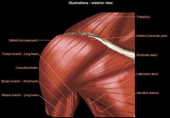
Posterior view of the shoulder girdle bones - Netter ... from s-media-cache-ak0.pinimg.com The rotator cuff is a made up of four muscles in the shoulder, connecting the humerus to the scapula. The muscle of the anterior compartment (arm in anatomical position) function as flexors while the muscles of the posterior compartment function as extensors. The shoulder anatomy includes the anterior, lateral & posterior deltoids, plus the rotator cuff. Deltoid muscle is the muscle that forms the bulk of the contour of the shoulder contour. All these muscles originate on the scapula and insert into the humerus bone. Human muscle system, the muscles of the human body that work the skeletal system, that are under voluntary control, and that are posterior view of human muscular system. This flow diagram provides an aid to diagnosis of shoulder conditions They are also categorized figure 1:
This muscle diagram is interactive:
These smaller muscles help to move substances through the body and support the function of these organs and vessels. They are also categorized figure 1: The muscle of the anterior compartment (arm in anatomical position) function as flexors while the muscles of the posterior compartment function as extensors. Posterior band of the ighl. Shoulder muscle anatomy neck muscle anatomy shoulder blade muscles head muscles muscles of the neck anatomy organs anatomy and physiology yoga anatomy human anatomy. Muscles of the shoulder can be divided into two strata: The shoulder muscles are associated with movements of the upper limb. The posterior muscles of the shoulder: The posterior view of the arm with the supraspinatus, infraspinatus, teres minor, and teres major rotator cuff muscles of the shoulder. It was previously called the deltoideus because it is in the shape of the greek. Case contributed by mr gray's illustrations. Thought consistent with impingement syndrome. This muscle diagram is interactive:
Click on the name of a muscle for a page about that muscle (works for most labels). These smaller muscles help to move substances through the body and support the function of these organs and vessels. Muscle length assessment edit source. Flexes and medially rotates arm; The scapula (shoulder blade) is elevated by the trapezius muscle , which runs from the back of the neck to the middle of the.
Muscles of the Neck and Torso - Classic Human Anatomy in ... from doctorlib.info Anterior part of the deltoid: The scapula (shoulder blade) is elevated by the trapezius muscle , which runs from the back of the neck to the middle of the. Pain in the shoulder joint. Related posts of shoulder muscles labelled diagram. Posterior muscles of the body diagram (with images). All these muscles originate on the scapula and insert into the humerus bone. • coracobrachialis • pectoralis major • subscapularis. Summary of the structure of the posterior shoulder muscles.
Thought consistent with impingement syndrome.
Posterior muscles of the arm and forearm. The anterior, lateral and posterior deltoid heads. Infraspinatus and teres minor tendon. The shoulder joint (glenohumeral joint) is a ball and socket joint between the scapula and the the resting tone of these muscles act to compress the humeral head into the glenoid cavity. Want to learn more about it? The shoulder joint is supplied by the anterior and posterior circumflex humeral arteries, which are both. Case contributed by mr gray's illustrations. Extends and laterally rotates the arm. The shoulder muscles are associated with movements of the upper limb. Anterior graphic of the shoulder. Learn vocabulary, terms and more with flashcards, games and other study tools. Unidirectional posterior shoulder instability is much less common than anterior instability, however it should be strongly suspected in those high risk group of athletes with posteroir shoulder pain and/or clicking. They are also categorized figure 1:
Posterior shoulder pain is more often than not mistakenly identied as rotator cuff disease or cervical disk disease. Case contributed by mr gray's illustrations. This muscle diagram is interactive: Muscle length assessment edit source. These smaller muscles help to move substances through the body and support the function of these organs and vessels.
Upper body weight training exercises from staticg.sportskeeda.com Unidirectional posterior shoulder instability is much less common than anterior instability, however it should be strongly suspected in those high risk group of athletes with posteroir shoulder pain and/or clicking. The human shoulder is made up of three bones: This page is about human muscle diagram posterior,contains hb muscular system posterior,human muscle system functions, diagram, & facts,anterior muscle diagram anterior muscle diagram. The anterior, lateral and posterior deltoid heads. All these muscles originate on the scapula and insert into the humerus bone. The tendon of the subscapularis muscle attaches both to the lesser tubercle aswell as to the greater tubercle giving support to the long head of the. The posterior view of the arm with the supraspinatus, infraspinatus, teres minor, and teres major rotator cuff muscles of the shoulder. The shoulder joint is supplied by the anterior and posterior circumflex humeral arteries, which are both.
Thought consistent with impingement syndrome.
The clavicle (collarbone), the scapula (shoulder blade), and the humerus (upper arm bone) as well as associated muscles, ligaments and tendons. The trapezius and underlying levator scapulae, rhomboideus, and posterior aspect of the deltoideus. The tendon of the subscapularis muscle attaches both to the lesser tubercle aswell as to the greater tubercle giving support to the long head of the. Posterior part of the deltoid: The human shoulder is made up of three bones: This flow diagram provides an aid to diagnosis of shoulder conditions Want to learn more about it? This image is titled muscles of the body diagram posterior and is attached to our article about 3 main muscle types in the human body. Nine muscles cross the shoulder joint. Posterior muscles of the body diagram (with images). • coracobrachialis • pectoralis major • subscapularis. Pain in the shoulder joint. Patients with muscle tenderness are diagnosed with myofascial pain. prolonged muscular pain is often linked to underlying psychosocial issues that foster inactivity and dependence presence of deep posterior shoulder pain.
All these muscles originate on the scapula and insert into the humerus bone shoulder muscles diagram. Posterior muscles of the arm and forearm.


Posting Komentar
0 Komentar